

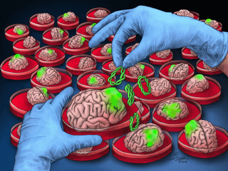
In a new study published in Molecular Psychiatry, researchers from Sweden and a Harvard-affiliated hospital in Boston tried to find out by creating “brain organoids,” or miniature brains about the size of a pinhead, and infected them with COVID.
What they saw explained a lot: An “excessive number” of synapses, or connections between brain cells that allow them to communicate, were eliminated during the course of the disease—“more than you would expect to see in a normal brain,” the authors wrote in an Oct. 27 article on academic news site The Conversation.
In a process known as “pruning,” normal brains get rid of a certain amount of inactive synapses when they’re no longer needed, to make way for new ones. But the infected mini brains showed unnecessary and inordinate levels of the clean-up process, similar to the level seen in neurological disorders like schizophrenia, Alzheimer’s, and Parkinson’s disease, according to the authors.
Authors of the new study caution that because the mini brains they created are so small, they may resemble the brain of a fetus more than that of an adult. Still, some studies of individuals who’ve died from COVID, in addition to imaging studies of those who’ve survived it, report the death of neurons and a reduction in the thickness of gray matter in the brain, signs of synapse loss, they say.