Imagine that you are in a position to buy an amazing invention–a new robot. It’s top of the line, able to perform numerous, complex functions, both physical and mental. But there’s a catch. You must supply it with power constantly, never letting the battery discharge down to zero. Should that happen and you attempt to reload it with power, the robot explodes. It’s a serious design flaw, and probably most tech-savvy people would think twice before buying such a machine. But it’s a close analogy to what destroys a human brain and other vital organs of the body.
When the heart stops beating, cutting off blood flow to the brain, when oxygen is depleted from the blood, or when something prevents oxygen from moving out of blood cells and into body tissues where it’s needed, the brain dies, but not immediately. Leave body cells in culture medium in a laboratory without oxygen and the cells appear almost normal. They’re dormant—not active but existing in a kind of sleeping state—until the oxygen supply is restored and that’s when a cascade of biochemical events begins that actually leads them to die.
The same thing happens with the cells of a living brain after a person who has been in cardiac arrest—lacking a heart beat—for a few minutes of longer has their heartbeat restored. Blood flow brings oxygen into the oxygen-starved brain and that’s what actually causes brain damage and death. It’s called re-perfusion injury and it’s why a person whose heart has stopped often can have their heart restarted fairly easily, but then later (typically about 24 hours after the heartbeat has been restored) they die.
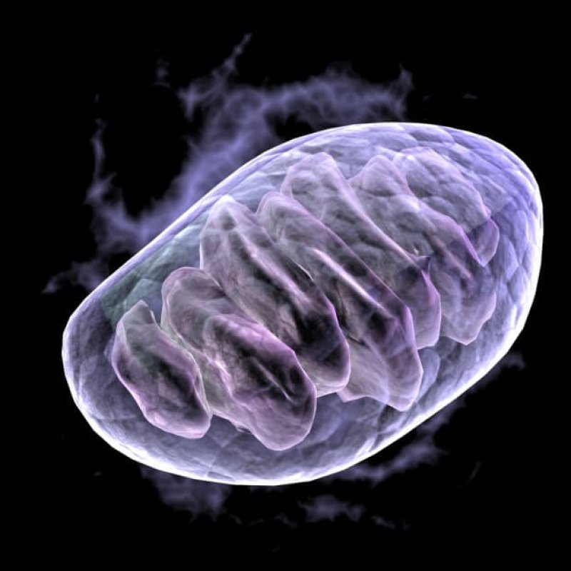
We’re not going to build robots with this type of design flaw, and when it comes to humans the good news is that we’re honing in on what causes the problem in our own cells. It’s due to cellular organelles called mitochondria, the power plants of the cell, where oxygen is used to draw energy out of food molecules in quantities much higher than can be drawn out without oxygen. Along with understanding the role of mitochondria in re-perfusion injury comes the potential for using the knowledge in clinical applications.
That’s the part that’s particularly exciting, because the applications are already leading to a scenario in which the amount of time between cessation of the heartbeat and irreversible death—a time that used to be limited to four minutes—is being extended, and not just in a hypothetical way, but in hard guidelines being used in many hospitals. That’s true already when it comes to one intervention—therapeutic hypothermia, the intentional lowering of a patient’s body temperature—and it’s on the verge of becoming reality as scientists learn more about how mitochondria function and how we may take control of them.
When mitochondria are extracted from cells of humans or other organisms, ground up and injected into an animal under sterile conditions, they cause an inflammatory reaction throughout the body. This is the same thing that happens when animals including humans, are infected with certain bacteria. That’s because the mitochondria in our cells are actually bacteria themselves. Mitochondria have their own genes and genomic studies show that the ancestors of mitochondria were incorporated into the ancestors of our cells about 2 billion years ago. Mitochondrial ancestors were a kind of bacteria—called aerobic bacteria—that could use the oxygen that was building up in large quantities in Earth’s atmosphere and oceans. By taking in aerobic bacteria, our single-celled ancestors gained the ability to generate large amounts of energy using oxygen, and by residing within the much larger cells the bacterial gained protection from the outside environment.
Mitochondria have in the news because they have retained many of their own genes that can be used to track the maternal lineage of humans. That’s because we get them only from our mothers, and this phenomenon has also made possible the three-parent baby, which has also enjoyed wide media coverage, due to the many social issues that it raises. There are literally hundreds of different medical conditions caused by various flaws and some of these have been in the news too.
From a genetic standpoint, it’s hard to assess the incidence of most of these mitochondrial diseases, because the penetrance—how influential a genetic problem actually ends up being in a person– varies widely. Also, some mitochondrial diseases depend on both parents, because they result from inherited genes that are not present in the mitochondria themselves, but in the cell nucleus, yet many mitochondrial diseases come from mutations occurring within the egg that gives rise to the individual. These conditions are not passed down through generations, not even from the mother, since they occurred only in some of the mothers eggs, rather than in all of her cells. But accumulating evidence supports a growing consensus that flawed mitochondrial function may underly some of the most common conditions, such as diabetes, cancer, and obesity –particularly, an obesity-related syndrome known as metabolic syndrome.
Extending the time to for resuscitation
By far, the leading cause of death is heart disease and it turns out that mitochondria also play a major role in this category of disease. Fortunately, most myocardial infarctions (MIs; “heart attacks”) show their effects over a long-enough time for victims to be treated before their hearts are damaged to such an extent that the pumping action stops. In such cases, the treatment is often successful and the person goes on to live for many years. But if the heart actually does stop, meaning they going into cardiac arrest, the prognosis is much worse. Their blood pressure drops to zero and they lose consciousness. In many such cases, the heart stops in a way that it can be restarted and the blood pressure restored, but resuscitation alone is not enough to save them in most cases. Because of the re-perfusion, the brain is damaged and the chances of damage are high, particularly if the victim is in a coma following resuscitation.
The thinking today is that a chain of enzymes within cell mitochondria, known as the electron transport chain (ETC), is inundated with oxygen and with the various molecules from which the oxygen is normally used to draw energy. This causes massive production of entities called oxygen free radicals, which ultimately signal the cell to commit suicide.
For the last decade, however, the American Heart Association has had guidelines in place for reducing re-perfusion injury using therapeutic hypothermia. After resuscitation, patients are put into an intentional coma, a cold coma that’s induced by cooling their bodies a few degrees (most protocols use 32 to 34 degrees Centigrade as the target temperature) and using drugs to prevent shivering. They’re kept at low temperature for 24 hours and since the guidelines have been in place, survival after cardiac arrest has more than doubled in hospitals where the technique is performed. The protocols are constantly being tweaked with the goal of improving survival and the message of the relevant scientific research is that it works because the low temperature tempers the reactivation of the ETC within mitochondria.
What’s possibly even more exciting is the discovery in recent years that various chemicals that are usually poisons — horrible compounds like hydrogen sulfide, carbon monoxide, and hydrogen cyanide, which are known to paralyze the mitochondrial ETC—actually are made within our cells in small quantities. This has stimulated an emerging idea that such poisons actually are needed in small quantities for mitochondrial regulation and that it might be possible to turn off mitochondria and turn them back on again without triggering the cells containing them to commit suicide.
Initially, it may sound like pie in the sky, but using hydrogen sulfide, Mark Roth of the Fred Hutchinson Cancer Center in Seattle has been putting mice and other small mammals into suspended animation—a dormant state with complete cessation of metabolic activity — and then waking them up with no ill effects. Roth and his team has been doing this for more than 15 years and has also been working with larger animals like pigs with promising results.
We’re only at the beginning of learning how to use all of this new knowledge, but we seem to be on course. And it’s all due to those tiny little organelles that 2 billion years ago just needed a place to live.
David Warmflash is an astrobiologist, physician and science writer. Follow @CosmicEvolution to read what he is saying on Twitter.

