

The GLP aggregated and excerpted this blog/article to reflect the diversity of news, opinion and analysis.
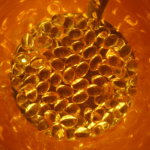
Johns Hopkins researchers have developed a method to efficiently turn human stem cells into retinal ganglion cells, the type of nerve cells located within the retina that transmit visual signals from the eye to the brain. Death and dysfunction of these cells cause vision loss in conditions like glaucoma and multiple sclerosis.
“Our work could lead not only to a better understanding of the biology of the optic nerve, but also to a cell-based human model that could be used to discover drugs that stop or treat blinding conditions,” says study leader Donald Zack, M.D., Ph.D., the Guerrieri Family Professor of Ophthalmology at the Johns Hopkins University School of Medicine. “And, eventually it could lead to the development of cell transplant therapies that restore vision in patients with glaucoma and MS.”
The laboratory process, described in the journal Scientific Reports, entails genetically modifying a line of human embryonic stem cells to become fluorescent upon their differentiation to retinal ganglion cells, and then using that cell line for development of new differentiation methods and characterization of the resulting cells.
Using CRISPR-Cas9, investigators inserted a fluorescent protein gene into the stem cells’ DNA. This red fluorescent protein would be expressed only if another gene was also expressed, a gene named BRN3B (POU4F2). BRN3B is expressed by mature retinal ganglion cells, so once a cell differentiated into a retinal ganglion cell, it would appear red under a microscope.
Read full, original post: Growing Retinal Nerve Cells in a Lab