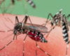


Even something as tiny as a cell is thick enough for specialized cameras to examine in detail. In a process called Z stacking, scientists photograph their subject, whether it be a cell or bit of pollen, and choose a series of different focal points for each picture. When viewed sequentially, the images illustrate the contents and contours of the subject.
Compiled and edited by Dylan T. Burnette and Aidan M. Fenix, both at the Vanderbilt University School of Medicine, this GIF highlights several cell components. The egg-shaped silhouette is the nucleus, or control center, which encases DNA. The vertical marks on the left of the nucleus are the Golgi apparatus, the structure that packages proteins and sends them to their destination. Crisscrossing and encircling the entire cell are protein threads. Some are chains of a single protein, while others bundle two molecules together. These strands maintain the cell exterior and contract it into new shapes.