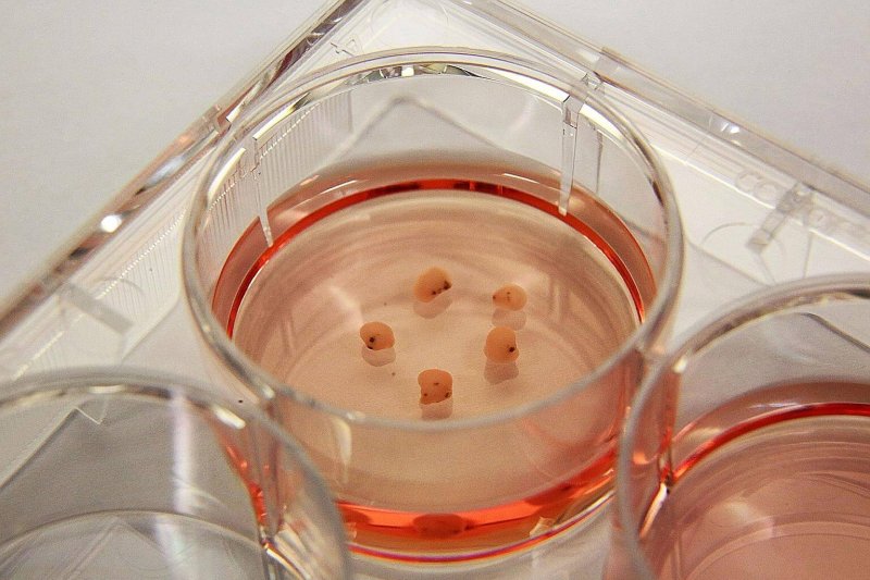
The fact that they’re easy to make and behave a lot like the organs they resemble make them an exciting tool in the scientific study of health and disease. And, while they are still in their infancy in terms of development, organoids have a lot of potential in investigating the cause of neurodevelopmental disorders that haven’t yet been cracked using more well-established methods, such as studying mice or two-dimensional cell cultures.
We have mice to thank for much of what we know about neurodevelopmental diseases. You may not think you have much in common with a mouse, besides a love of cheese, but we are quite similar at the genetic level. When common genes get messed up in mice, they end up with diseases that look similar to the way they do in people. However, this type of study doesn’t work as well when looking at diseases that are caused by more than one genetic mutation – diseases like schizophrenia or autism, which are caused by multiple genes, are almost impossible to fully model in mice.
 Enter organoids. Made out of stem cells, in their first incarnation (back in 2001) scientists figured out how to turn them into neural progenitor cells, self-renewing cells that can develop into the cells that make up the brain, like neurons and glia, but are not yet committed to one fate. These neural progenitor cells self-organized in 2D structures called rosettes that displayed features of the embryonic neural tube, the precursor to the nervous system. Ten years later, scientists found a way to go a step further: it became possible to create structures that look somewhat like the developed brain as a whole – or distinct regions of the developed brain, which could be fused together.
Making organoids
To make these 3D structures, you basically have to recreate the same environment within which the brain develops. We don’t fully understand all the processes involved in neurodevelopment but, based on what we do know, it has been possible to recreate some important aspects of the process. For example, the protein SMAD, which is responsible for muscle and skin formation, can be inhibited, while the WNT signaling pathway, which involves numerous molecules responsible for regional patterning, can be carefully controlled, giving rise to different anatomical brain parts.
With that technological know-how, and the ability to turn a patient’s cells back into pluripotent stem cells, it’s possible to turn cells from patients who have complex diseases like schizophrenia into brain organoids, in a dish. We’ve been reprogramming and culturing patient-derived cells into pluripotent stem cells – with the kind of idiosyncratic genetic composition that is true to the nature of the genetic basis of schizophrenia – for years. But the ability to do this together with organoid technology is a step forward.
New models for psychiatric study
This technology provides an incredible opportunity for us to better model diseases, as the more complex tissue structure offers new possibilities for cellular and molecular analysis. It hasn’t been extensively used to study neuropsychiatric disorders yet. But the few studies that have been done, like one in which organoids were generated from four patients with Austism Spectrum Disorder to reveal genetic abnormalities, are promising. Scientists speculate that self-patterning 3D brain organoids will allow for more accurate study of the cell-to-cell interactions that are involved in the formation of synapses, myelination (wrapping up nerve cells in a fatty white substance, which is crucial for proper signaling) and circuit maturation, all of which have been implicated in schizophrenia.
It’s not just psychiatric diseases that could be better understood with the help of organoids. They are also being used to investigate the underpinnings of neurodevelopmental disorders. Because organoids resemble early stages of development, they have a lot of potential for studying developmental diseases that manifest in early embryonic fetal stages.
 In fact, organoids have been useful following the outbreak of Zika virus in 2016. Zika caused birth defects in babies born to infected pregnant mothers, including microencephaly, where babies are born with smaller-than-average heads and brain damage. Using organoids, it was possible to identify a causal relationship between Zika infection and the destruction of neural progenitor cells; one study showed that organoids exposed to Zika underwent growth reduction, consistent with the observation of microencephaly in affected infants. Another reported reductions in the number of neural progenitor cells and neurons as a result of cell death.
Organoids are also being used to explore the mechanisms behind human cortical expansion and surface folding, which give brains their funny convoluted, appearance and cannot be properly explored in mice, which have smooth, simple brains. Gyrification, the process of forming folds, can be partially achieved in human brain organoids.
The biggest question facing researchers when it comes to organoids is how well they model human development. The answer, at present, is: not quite. But they are getting better.
Limits of mini-brains
The brain continues to develop after birth. While organoid development resembles the prenatal period quite well, many aspects of brain development, including the maturation of cortical circuits, takes years to develop in growing humans, making it impractical to model these processes in organoids. There also remains much work to be done in terms of achieving more complex aspects of cortical organization, such as gyrification, establishing proper neural connections, and proper cortical layering.
On a more intricate level, genes are not expressed in exactly the same way in brain organoids as they are in human brains. The fact that the genetic identity of the different cell populations in brain organoids do not perfectly reflect the genetic identity of the different cell populations in the human brain needs to be addressed before organoids can be considered an airtight way to research human diseases. The difficulty is that during development, genes are turned on and off in an extremely controlled manner, both temporary and spatially, to create the brain as we know it, and these patterns are difficult to achieve artificially, because the complicated processes involved are not yet fully understood.
Despite their current limitations, organoids represent the next generation in artificial cell-based models of the human brain. These “mini-brains”offer a complex system for studying the neurobiology of countless brain disorders – in a dish. Over the coming years, more and more scientists are likely to adopt these 3D mini-brains, expanding their application and in the process giving rise to improvements in current methods.
I cannot wait to see where the field goes. I work on a stem cell project which aims to cure a rare, genetic childhood neurobiological diseases called Sanfilippo syndrome Type B by supplying the brain, which lacks a particular protein, with neural stem cells that can provide a long-term supply of that protein. These neural stem cells have the potential to differentiate into different neuronal cell types. By injecting organoids into the brains of experimental models, it may be possible to provide the brain with a useful combination of already-differentiated cell types, increasing the therapeutic potential of our stem cell therapy. Not only are mini-brains helping scientists find answers to intractable genetic questions – they may also help to solve them.
Yewande Pearse is a Research Fellow based at LA Biomed. She is working on stem cell gene therapy using CRISPR-Cas9 to treat Sanfilippo Syndrome.
A version of this article was originally published at Massive as “One great way to study brain diseases? ‘Mini-brains’ grown in dishes” and has been republished here with permission.

