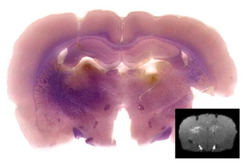
Typically, when scientists and doctors use MRI (magnetic resonance imaging) to examine the brain, they’re looking at blood flow and using it as a proxy for brain activity. Increased blood flow to a particular region of the brain is indicative of increased activity.
A team at MIT, led by biological engineer Alan Jasanoff, has come up with a trick using genetic engineering to make gene activity in the brain visible in living organisms using the same MRI that is already in wide use. This could be useful in understanding the roots of how our genes influence our ability to form memories and learn skills.
Anne Trafton from MIT News reports:
“The dream of molecular imaging is to provide information about the biology of intact organisms, at the molecule level,” says Jasanoff, who is also an associate member of MIT’s McGovern Institute for Brain Research. “The goal is to not have to chop up the brain, but instead to actually see things that are happening inside.”
Jasanoff and his team did so by creating an artificial “reporter gene” and testing it in mice. It’s a two-step process. First, they designed the gene and incorporated it into the brain cells of mice via a virus, which took up the gene and began producing an enzyme called SEAP.
Then, they injected a a magnetic contrast agent into the mice’s brains — without killing or harming them. Normally, the contrast agent dissolves and is invisible to the MRI, but in the presence of the SEAP enzyme from the reporter gene it accumulates and becomes visible to MRI. In other words, wherever in the brain the reporter gene was being expressed, the researchers were able to detect it via contrast agent and MRI.
This was a proof-of-concept experiment, so the team only used MRI to see if their reporter gene was successfully taken up my mice brains and interacted with the contrast agent the way they hoped. It did. The next step is to custom-design different versions of the SEAP reporter genes that will only activate when certain other genes are on, revealing the activity of these genes via MRI.
This novel approach to studying genetic activity in the brain recieved high praise from other scientists:
Assaf Gilad, an assistant professor of radiology at Johns Hopkins University, says the MIT team has taken a “very creative approach” to developing noninvasive, real-time imaging of gene activity. “These kinds of genetically engineered reporters have the potential to revolutionize our understanding of many biological processes.”
The thought of needing an inter-cranial injection to enable a genetic-level brain scan may sound invasive, but until now the only way to get at genetic activity in brains was by taking them out and slicing them up; if the MIT team’s technique works as intended it could open up a new window into how our genes and brains interact.
- Read Anne Trafton’s full article at MIT News: “MRI reveals genetic activity“
- Read the original paper in Cell Biology: “MRI-Based Detection of Alkaline Phosphatase Gene Reporter Activity Using a Porphyrin Solubility Switch“
Kenrick Vezina is Gene-ius Editor for the Genetic Literacy Project and a freelance science writer, educator, and amateur naturalist based in the Greater Boston area.
Additional Resources:
- “Injecting DNA in the brain: What’s the promise of gene therapy for Parkinson’s disease?” Tabitha M. Powledge | Genetic Literacy Project
- “Brain science pioneer shifts focus of NIMH to basic neuroscience and genetics,” Benedict Carey | New York Times
- “Does reading a novel physically change your brain?” Jeremy Summers | Genetic Literacy Project


